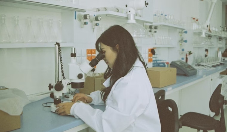
Scientists at The Ohio State University are making strides in understanding the intricate communication between cells, using tiny bubbles known as extracellular vesicles and particles. According to Ohio State News, these microscopic messengers play a role in human health and disease. However, the specific contents and their impacts on recipient cells largely remain under wraps.
Breaking new ground in this field, a study published in Nature Methods details a novel approach by Ohio State researchers to immobilize these cellular couriers. This is significant because, as the leader of the study, Eduardo Reátegui explained to Ohio State News, "The extracellular vesicles present in tissues are very poorly understood in terms of how the particles actually interact with cells in our body." By immobilizing the vesicles, the team hopes to unravel the mystery surrounding their functions and origins.
The procedure, dubbed Light-Induced Extracellular Vesicle and Particle Adsorption (LEVA), doesn't harm the vesicles but instead allows them to be studied individually or in clusters. The process begins with a chemically coated glass surface, upon which UV light is used to create micropatterns that attract the proteins on the vesicles' exteriors through electrostatic charge. This method was shown to be effective in experiments, ensuring vesicles adhered strictly to the illuminated patterns.
Not only does LEVA advance research capabilities, but it could also revolutionize early disease diagnosis and drug delivery methods. "What we want to do is not only understand what these vesicles contain, but also identify their tissue of origin, and how they interact with cells," Reátegui told Ohio State News. Prior attempts to analyze these particles involved antibodies, limiting the researchers to only those vesicles matching a specific molecule. LEVA, however, enables unbiased immobilization of all vesicles, providing a fuller picture of their role in bodily functions.
An instance of the study's practical application showed LEVA's potential for examining immune responses. Researchers mimicked the behavior of immune cells to bacterial extracellular vesicles and observed what is known as a neutrophil swarming. "This showed that, first, we can generate matrix-bound EVPs in different contexts for easy analysis, and second, this approach allows exploration of EVP interactions in tissue," Reátegui said to Ohio State News.
Contributions to this research came from across academic disciplines, with funding from bodies like the Ohio State Center for Cancer Engineering-Curing Cancer Through Research in Engineering Sciences, the National Institutes of Health, the Burroughs Wellcome Fund, and postdoctoral scholar programs at Ohio State. The implications of these findings could touch numerous aspects of medical science, providing insights into the fundamental processes of cell-to-cell communication.