MIT and Brigham and Women's Hospital researchers have struck new ground in brain tumor diagnosis with a revolutionary microscopy technique that reveals details in human brain tissue like never before. Their findings, which highlight the potential for aggressive cells in low-grade tumors, could be a game-changer for how brain tumors are understood and treated.
In a groundbreaking study, scientists detailed how the new imaging method has exposed cell types and structures previously hidden to the human eye. According to a report by MIT News, this expansion microscopy technique involves physically enlarging the tissue samples, thus allowing the use of standard light microscopes to produce high-resolution images. This method could revolutionize not just the diagnosis, but also the prognosis and therapy choices.
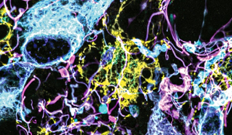
"We’re starting to see how important the interactions of neurons and synapses with the surrounding brain are to the growth and progression of tumors. A lot of those things we really couldn’t see with conventional tools, but now we have a tool to look at those tissues at the nanoscale and try to understand these interactions," explained Pablo Valdes, former MIT postdoc and the study's lead author, in an interview reported by MIT News.
The research, published in Science Translational Medicine, was conducted under the guidance of senior authors Edward Boyden from MIT and E. Antonio Chiocca from Harvard Medical School. The expansion microscopy technique leverages a softening protocol, allowing antibodies to label proteins after tissue expansion. "We open up the space between the proteins so that we can get antibodies into crowded space that we couldn’t otherwise," Valdes said, noting the technique's efficiency in protein labeling and imaging in dense tissue samples.
The team tackled challenging human brain tissue samples, which are typically embedded in paraffin and require extensive preparation. Their efforts yielded the visualization of up to 16 different molecules per sample, marking a significant step forward in the understanding of conditions like gliomas. To the researchers' surprise, "low-grade" gliomas displayed a concerning abundance of vimentin-expressing tumor cells, which are indicative of high aggressiveness in glioblastomas. "This tells us something about the biology of these tumors, specifically, how some of them probably have a more aggressive nature than you would suspect by doing standard staining techniques," Valdes stated, as per the MIT News report.
Chiocca emphasized the potential impact of this discovery on patient care, "These are incurable brain cancers, and this type of discovery will allow us to figure out which cancer molecules to target so we can design better treatments.” With a focus on creating a diagnostic tool for practical use in neuro-oncology, the researchers look forward to broadening their analysis of tumor types to develop solid diagnostic guidelines, as mentioned by the MIT News report.
The collaborative research found not only fundamental support from the academic community but also received funding from notable entities like the Howard Hughes Medical Institute, the Bill and Melinda Gates Foundation, and the National Institutes of Health. As Boyden's lab moves forward to explore other brain function aspects, the study signifies a substantial leap for nanoimaging and its role in the ongoing battle against brain cancer.