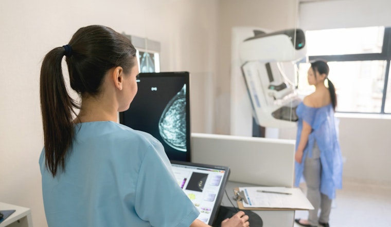
In a promising development for breast cancer diagnosis, researchers from MIT and ETH Zurich have introduced an AI model that can distinguish between various stages of a common preinvasive tumor. Serving as a potential tool to reduce overtreatment of ductal carcinoma in situ (DCIS), which can occasionally evolve into invasive cancer, the model leverages readily available breast tissue images. The simplicity of this novel AI approach, detailed in a recent publication in Nature Communications, proposes a significant step in the staging of DCIS.
Accounting for around 25 percent of all breast cancer cases, DCIS has posed challenges for clinicians due to the difficulty in pinpointing the precise nature and progression of the condition. The newly developed AI model by the team reportedly distinguishes the stages of DCIS by examining both the condition and the arrangement of cells within a tissue sample, emphasizing a shift in strategies for dealing with the disease, a detail explained in the researchers' findings.
The model's database, comprising 560 tissue images sourced from 122 patients, underpins the AI's capacity to identify certain characteristic cell states relevant to DCIS. This critical pivot to utilizing machine learning could obsolete more laborious and expensive diagnostic methods, with Caroline Uhler, a professor at MIT, underscoring the scalability of the technology. "We took the first step in understanding that we should be looking at the spatial organization of cells when diagnosing DCIS," Uhler told MIT News. Her hope is to advance this method into prospective studies and ultimately clinical practice.
Collaboration was key in forming the research basis, which borrowed techniques such as a single chromatin stain that showed similar efficacy to the more costly single-cell RNA sequencing. The researchers designed a machine-learning algorithm that factored in not just the presence but also the positional relationship of cells indicative of invasive cancer, revealing that organizing these cells matters in the samples, as GV Shivashankar, from ETH Zurich, noted. "But in cancer, the organization of cells also changes," he said during the recent study, according to MIT News. These insights have enabled the AI model's accuracy to significantly increase, bringing new perspectives on cancer diagnosis.
Upon comparison with pathologist evaluations, the model's predictions often concurred with professional diagnostic outcomes, highlighting its potential as a supplementary diagnostic tool. Additionally, the technology's adaptability holds promise for other cancer types and neurodegenerative disorders. The multifaceted benefits of this research were made possible through support from institutions such as the Broad Institute, the Swiss National Science Foundation, and the U.S. National Institutes of Health, among others. The findings, as mentioned in an MIT News release, could usher in an era of AI-augmented precision in the fight against one of the most common cancers affecting women worldwide.