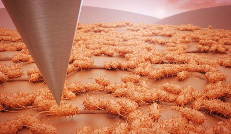
Scientists at Oak Ridge National Laboratory (ORNL) have radically improved the study of bacterial biofilms by enhancing atomic force microscopy (AFM). This new approach links the fine details of individual bacterial features with the grand scale of biofilm organization, providing clearer insights into these complex microbial communities. The groundbreaking research utilizes a large-area AFM platform developed by the lab's Center for Nanophase Materials Sciences, a Department of Energy (DOE) Office of Science user facility, as reported by the ORNL news release.
As a reminder of the pervasive influence of biofilms, they can incite infections, block pipes, and even disrupt environmental systems. Understanding them has significant implications across various sectors. The novel AFM platform contextualizes the positional details of individual bacteria within the broader panorama of biofilm topography. Despite its existing prowess in capturing intricate biological features, traditional AFM was hampered by its limited scope, which the new development at ORNL has now addressed.
Key to this advancement is the integration of machine learning with the AFM imaging process, enabling researchers to dissect and interpret copious amounts of data. According to ORNL's Dr. Sita Sirisha Madugula, a postdoctoral researcher and co-author, this methodology facilitated the automatic analysis of over 19,000 bacterial cells. "The integration of machine learning allows us to extract meaningful quantitative data from these massive datasets." The resulting biofilm maps reveal patterns resembling honeycomb structures and detail the role of flagella in the spatial organization and potential adaptation processes of biofilms, per the Oak Ridge National Laboratory.
The ORNL team also ventured to test the impact of engineered surfaces with nanoscale features on biofilm development, discovering that certain patterns could hinder traditional biofilm growth. This could lead to novel designs for surfaces that naturally repel bacterial colonization. Published in the journal npj Biofilms and Microbiomes, the research, according to Ruben Millan-Solsona, a postdoctoral researcher in ORNL's Functional Atomic Force Microscopy group and study co-leader, "Using the AFM, we could examine individual bacterial cells in detail but not how they organize and interact as communities." Co-leader and R&D researcher Liam Collins added, per the ORNL news release, "This new platform changes that. Now, we can visualize both the intricate structures of single cells and the larger patterns across entire biofilms."