Researchers at MIT have created a wearable ultrasound monitor that can gauge urinary bladder fullness without an ultrasound operator or application of gel. According to the MIT News, the product stands as a potential aid in monitoring and diagnosing bladder and kidney disorders, as well as deep tissue cancers such as ovarian cancer.
The revolutionary ultrasound patch accurately images the bladder. Possessing the potential accurately to track these organs' function, the technology could be adapted to probe other deep body tissues. Consequently, this advancement may enable earlier detection of deep-seated cancers, stated MIT professor Canan Dagdeviren.
The design of this ultrasound patch was partly motivated by Dagdeviren's younger brother's struggle with kidney cancer and subsequent difficulty in emptying his bladder completely. "Millions of people are suffering from bladder dysfunction and related diseases, and not surprisingly, bladder volume monitoring is an effective way to assess your kidney health and wellness," as mentioned by Dagdeviren in the MIT News report.
Traditional ultrasound probes, bulky and necessitating a medical visit, currently carry out bladder volume monitoring. Aiming for making this monitoring accessible at home, the MIT research team has developed a wearable alternative.
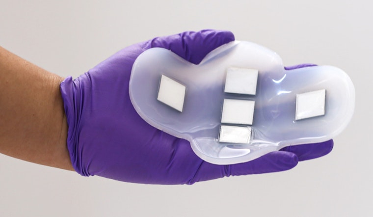
The patch is a flexible structure crafted of silicone rubber, housing five ultrasound arrays. These arrays, formed from a novel piezoelectric material, are organized in a cross shape. To be capable of imaging the entire bladder, which measures approximately 12 by 8 centimeters when full. The patch, adhering naturally to the skin through the polymer used, can be further secured by clothing.
A study conducted with collaborators at the Center for Ultrasound Research and Translation and Department of Radiology at Massachusetts General Hospital revealed that the patch could create images equivalent to those generated using a traditional ultrasound probe. The images, not requiring ultrasound gel or applied pressure, can serve to monitor changes in bladder volume.
Presently, the ultrasound arrays pair with a standard ultrasound machine found to image in medical centers, but the team envisions a portable device similar to a smartphone size for viewing the images. This advancement would enhance further the accessibility of bladder volume monitoring for patients.
The team of MIT aims to transform bladder and kidney health monitoring, and in pursuit of this aim, they plan the lofty goal of designing an ultrasound device that can image other organs like the pancreas, liver, or ovaries. To adjust the ultrasound signal's frequency based on each organ's location and depth, they need to develop new piezoelectric materials. For some deep organs, an implant rather than a wearable patch may yield better results.
Highlighting the substantial potential of this technology, Anantha Chandrakasan, dean of MIT's School of Engineering, stated that "this work could develop into a central area of focus in ultrasound research, motivate a new approach to future medical device designs, and lay the groundwork for many more fruitful collaborations between materials scientists, electrical engineers, and biomedical researchers," as reported by MIT News.