























































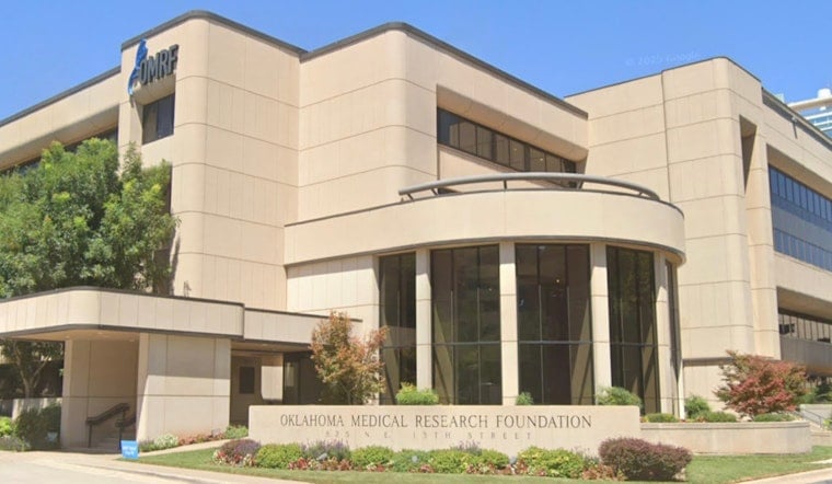
In a twist that could rewrite what doctors think they know about scars, researchers at the Oklahoma Medical Research Foundation in Oklahoma City say fully mature fat cells are not as settled down as everyone assumed. In mouse experiments, the team followed individual adipocytes during wound healing and found that some of these cells shed their lipid stores, started dividing and took on the hallmarks of scar-forming fibroblasts. Even after the wounds closed, the transformed cells kept their fibroblast-like identity, suggesting the switch is long-term rather than a brief detour.
The findings are detailed in Cell Reports, where the authors used intersectional lineage tracing to mark Adipoq+ adipocytes and track Pdgfra+ and Pdgfrb+ descendants in healing skin. The analysis turned up adipocyte-derived PDGFR+ cells (ADPCs) that cranked up collagen and smooth muscle actin while re-entering the cell cycle. The paper also reports that active PDGFRα signaling boosted ADPC proliferation and supported angiogenesis and re-epithelialization during repair.
How the Team Tracked Cells
According to OMRF, the experiments were carried out in mice and relied on genetic tracing over a three-week (21-day) window to shadow individual fat cells through the repair process, with sequencing data made publicly available. The study, led by Lorin Olson, Ph.D., used a two-step tracing strategy to separate each cell's adipocyte past from its newly gained fibroblast markers. The listing notes that funding came from the National Institute of Arthritis and Musculoskeletal and Skin Diseases (NIH), the Oklahoma Center for Adult Stem Cell Research (a TSET program) and the Presbyterian Health Foundation.
What Researchers Said
Courtney Griffin, Ph.D., OMRF's vice president of research, summed up the surprise by saying fat cells "are much more adaptable than we realized," as reported by The Journal Record. Olson told the outlet that "we've long viewed fat cells as having reached their final identity," and the team said they hope the work will eventually guide strategies to curb excessive scar tissue and surgical adhesions. The researchers also emphasized that once the wounds healed, the converted cells did not drift back to being fat but stayed locked in a fibroblast-like state.
Implications and Caveats
The authors argue that the discovery exposes a previously underappreciated source of scar-building cells and flags new molecular targets for managing fibrosis, but they stress that all of the evidence so far comes from mice. In the Cell Reports study, tweaking PDGFR signaling altered both how the cells behaved and how the wounds healed, a reminder that any future treatment would have to walk a fine line between limiting harmful scarring and preserving normal repair. The researchers caution that confirming the relevance to humans will require work in human tissues and thorough safety testing.
What's Next
The team has deposited sequencing data, with a GEO accession listed in the paper, and invited other labs to test whether the same adipocyte-to-fibroblast switch shows up in human tissue, according to OMRF. The Olson lab plans to keep dissecting the signaling cues that drive the transformation and to explore whether pharmacologic or genetic approaches can dial down damaging fibrosis without derailing healing. For now, the work recasts adipocytes as a surprisingly flexible cell population whose behavior during injury might be a practical target in anti-fibrotic therapies.
The finding challenges the long-held notion that fat cells are permanently locked into their role and steers researchers toward fresh angles on how scars form. As the science moves from mice into human tissue, OMRF investigators say the coming years will be key for learning whether this cellular flexibility can be turned into safe treatments for patients.